
Strabismus in children and adults. Causes and treatment
Strabismus, also known as “crossed-eyes” or “crossed vision”, is an ophthalmological disorder in which the visual axis of the eyes is not aligned. This causes one eye to deviate when it needs to look at a fixed point.
As a rule, this disease is observed from early life. Studies show that about 5% of children born in one year have strabismus, and 50% of them may suffer from low vision or lazy eye. Regular ophthalmological examinations are essential to detect and treat strabismus as far as possible.
What is strabismus?
Strabismus is a disease that affects binocular vision.
It can manifest:
- In one eye
- In both eyes
- Constant
- Intermittent
*Note: Although strabismus can only occur in one eye, complex processes in the brain areas often make the disorder visible and affect both eyes.
About eye muscles
The eye moves thanks to the 6 torsion muscles in the eyeball.
- 2 horizontal
- 2 vertical
- 2 obliques
They coordinate all eye movements as part of complex processes in the central nervous system. The functioning of the extraocular muscles is developed in the first months of life, and in order to orient the direction of gaze correctly, they must all be in absolute synchrony. Strabismus involves a lack of harmonious functioning between them.

What causes strabismus?
The causes of strabismus can be multiple. It usually occurs in childhood due to genetic abnormalities, but there is a risk that it can also develop in adulthood as a consequence of other disorders.
- In principle, strabismus is caused by heredity. In 80% of cases, strabismus is inherited if there is astigmatism, myopia or hypermetropia in the family. However, there are also cases where the disorder occurs in children even if there is no history of refractive errors, but there are other vision problems.
- Failure to develop stable connections in the brain is a cause of congenital strabismus or infantile strabismus.
- Diseases affecting the nervous system can cause strabismus.
- Cranial nerve paresis and paralysis
- Brain tumors
- Head trauma
- Neonatal neurological history, such as dysmaturity, prematurity, cerebral palsy, or meningitis.
- High diopters can also cause this disease.
- Big differences in diopters between the two eyes can lead to this.
- Severe myopia can cause “heavy eye syndrome” – a certain type of strabismus.
- High hypermetropia is mostly associated with convergent strabismus.
- Congenital changes in the orbital bones can also cause strabismus. One example is Crouzon disease.
- Trauma to orbital structures can lead to strabismus.
- So do orbital tumors
- Congenital abnormalities of the extraocular muscles, or infection and inflammation of the extraocular muscles can trigger strabismus.
- Thyroid diseases, such as Graves, or other autoimmune thyroid disorders.
- Autoimmune diseases: multiple sclerosis, mitochondrial disease, myasthenia gravis.
- Accidental injury to the extraocular muscles by injecting various substances into the eye socket. In this case, strabismus is accompanied by double vision after local anesthesia for various ophthalmological surgeries: glaucoma, retinal detachment, etc.
- Cataracts and unilateral ptosis, also known as “drooping eyelid”, can cause strabismus because they reduce the visual acuity of one or both eyes.
- Ageing may be another factor that favors strabismus.
- Long time spent in front of screens at close range.
How many types of strabismus are there?
Ophthalmic disorder can manifest itself in several ways.
- Depending on the orientation of the gaze, it can be:
- Convergent strabismus/esotropia, where the eye looks towards the nose (inward)
- Divergent strabismus/exotropia, where the eye looks towards the ear (outward)
- Hypertropia, where the eye is deviated upward
- Hypotropia, where the orientation is downward.
- Depending on intensity:
- Low Strabismus – in these cases, the disease is noticed when the patient is tired. The rest of the time, they manage to control it and their binocular vision functions optimally.
- High strabismus – here, the patient’s eyes are already out of alignment and suffer from diplopia (double binocular vision). The angle of deviation between the eyes means that the image is no longer perceived as single in the brain, resulting in overlapping.
- Depending on the age at which it appears:
- Infantile/congenital strabismus. It occurs in infants in the first 6 months of life.
- Acquired strabismus. It can occur at any time and is caused by one (or more) of the causes listed in the previous section. As a rule, it also becomes apparent in childhood, between the ages of 2 and 6.
- Depending on frequency:
- Constant strabismus
- Intermittent strabismus
- Other classifications:
- Monocular Strabismus VS. Alternating strabismus
- Paralytic Strabismus VS. Restrictive strabismus
Strabismus in children
The first 6 months of a baby’s life are perceived as a period of physiological instability, when it is considered normal for the infant’s eyes to deviate from their normal position. These cases of strabismus are intermittent and usually facing outward (divergent strabismus). After the age of 6 months, the infant’s gaze should remain stable and parallel. If not, specialist intervention is needed to treat the strabismus according to its severity.
The parent should constantly observe the child’s progress in this regard, as late detection of strabismus can lead to severe amblyopia (poor performance of one/both eyes). Moreover, “crossed-vision” can be a symptom of other diseases such as retinal or nerve damage, ocular tumors, congenital cataracts and malformations.
As a treatment for the children, there are various therapies or treatments such as VTS therapy and strabismus surgery. Following examination, surgery may be recommended until the infant is 24 months old. Thereafter, it may be repeated, as vision and extraocular muscles continue to develop until the age of 18.
Strabismus in adults
In adults, acquired strabismus can have different causes.
- They are usually pathological in nature – oculo-motor paralysis caused by neuro-muscular diseases, traumatic brain injury, ocular or intracranial tumors, or strokes.
- However, there is also a high risk of illness with ageing. This type of strabismus usually manifests itself by double vision. Because they have had normal binocular vision in the past, patients manage to ignore one of the images by closing one eye or looking for a position to overlap them.
- Adults may also experience residual strabismus (operated on but not completely corrected) or consecutive strabismus (which has changed its appearance after surgery).
There are several treatment options for adults suffering from strabismus.
- Low severity strabismus can be corrected with prism glasses.
- For small or intermittent deviations, botulinum toxin is used. It is also useful in cases of paralysis of the extraocular muscles, and if the patient has had several unsuccessful surgeries.
- If the patient is experiencing a severe form, more surgery may be needed, especially if the strabismus has occurred under the age of 1 year and is accompanied by severe amblyopia.
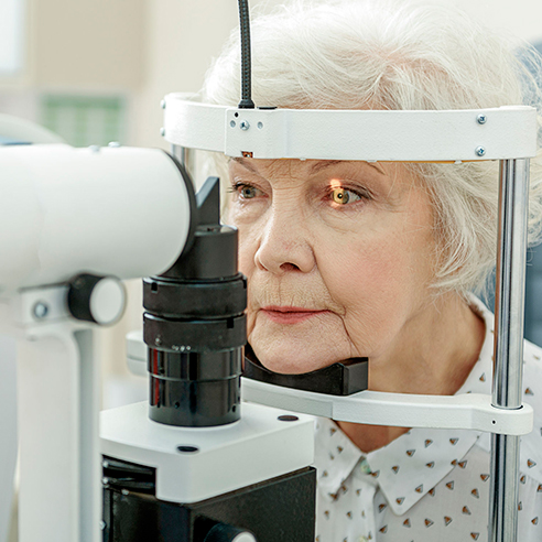
How is strabismus diagnosed?
It is a complex process that involves:
- Hereditary history check
- Examination of eyeball movements
- Eye opacity check
- Angle of deviation measurement
- Performing a prism test

How is strabismus treated?
How strabismus is treated differs, depending on the form of the disease and the age of the patient.
- If it is a mild form associated with a refractive error, glasses (with diopters corresponding to the refractive error) with prism lenses, which force the eye to stay straight, are recommended.
- In the case of strabismus and “lazy eye” syndrome, unilateral or alternate occlusion (to restore vision to the weak eye), wearing glasses and orthoptic exercises (to stimulate binocular vision; can be done in specialist clinics or at home under specialist guidance) are indicated.
- In severe forms, where the angle of deviation exceeds the physiological limit, surgical treatment is recommended.
If you are facing an ophthalmological problem, the specialists at Dr. Holhoș clinics are waiting for you with state-of-the-art technologies to provide you with the best care.

Nystagmus is the eye condition where the eyes make repetitive and uncontrollable movements. Discover other symptoms and treatment options.

Color vision deficiency, also known as dyschromatopsia, is a general term referring to various vision disorders characterized by a deficiency in color perception.

“Flying flies” are most often harmless and represent a normal stage in the aging process. Find out what the causes are and how you can reduce the symptoms.

Ocular allergies occur as a reaction of the body to an allergen, causing inflammation and itching in the eyes. The most common ocular allergies are seasonal.

Ophthalmic migraine is most common in the 40s. It manifests itself in visual impairment and even temporary blindness.

Keratitis, also known as “corneal ulcer”, is an inflammation of the cornea. If detected early, the ophthalmological disorder is easy to treat and heals quickly.
Diplopia is an ophthalmological disease in which you see two images of the same thing. The condition can affect anyone, but is more common after the age of 60.

Xanthelasma is a member of the xanthomas family and represents fatty deposits in the skin cells around the eyes. It is visible as yellow, harmless bumps.

Colorblind people perceive colors differently from most people. Most of the time, this ophthalmological disorder makes it difficult to distinguish between certain colors.

Epiphora is an ophthalmological disorder manifested by excessive tearing of the eyes. Most of the time, it is not severe and disappears on its own. However, if you are experiencing this and the problem persists, we recommend that you make an appointment for an ophthalmological examination. Treatment can be different, depending on the cause of the epiphora.

If you notice a yellow spot on the white of your eye, you are most likely dealing with pinguecula. The ophthalmological disorder is not severe, but the symptoms can be uncomfortable. Find out how to treat pinguecula and, more importantly, how you can prevent it.

Entropion is the ophthalmological disorder in which the eyelid of the eye turns inwards. It is different from ectropion, where the eyelid turns outwards. It most often occurs in older people and usually only affects the lower eyelid.

It is possible that you may also be experiencing ocular toxoplasmosis without knowing it. This retinal disorder is extremely common in our century and is manifested by eye discomfort and blurred vision.

Ectropion is the ophthalmic disorder in which the eyelid and eyelashes pull away from the cornea, and reorient outwards.

One of the most common types of headache is headache of ocular origin. It occurs as a result of an ophthalmological disorder.

Blepharitis is an ophthalmological disorder that manifests itself by inflammation of the eyelids. At the base of the eyelids, the patient notices small crusts formed by solidified oil particles or bacteria that collect in the crease at the corner of the eye.

Uveitis is an ophthalmological disorder of the uveal tract, manifested by changes in vision and eye pain.

Among the most common ophthalmological disorders is hordeolum. This is popularly known as an “stye” and is an infection of the eyelids.

The drooping eyelid is known in medical terms as “palpebral ptosis”. It manifests itself by narrowing the visual slit of one or both eyes, creating aesthetic and functional discomfort.

Amblyopia is a vision problem, popularly known as “lazy eye”. This disorder can occur in one or both eyes, and studies show that around 3% of the population suffer from this eye disease.

The sensation of “dry eyes” or “tired eyes” is known in medical terms as “dry keratoconjunctivitis” or “xerophthalmia”, and refers to a dysfunction of the tear film.

Strabismus, also known as “crossed-eyes” or “crossed vision”, is an ophthalmological disorder in which the visual axis of the eyes is not aligned. This causes one eye to deviate when it needs to look at a fixed point.

Conjunctivitis is one of the most common ophthalmological disorders. It can occur in adults, children and babies.

Chalazion is manifested by inflammation of the upper or lower eyelid. It is one of the most common ophthalmological disorders, and occurs when the secretion of sebaceous glands in the eye is blocked.

Macular degeneration involves deterioration of the macula and therefore of the quality of central vision. This disease does not affect peripheral vision and therefore cannot lead to complete blindness.
Hypermetropia affects the ability to see nearby objects. You may be able to see distant objects clearly, but closer objects, even words in a book, are usually out of focus. Hypermetropia occurs when the eye does not focus light properly on the retina (the light-sensitive layer at the back of the eye).

Myopia is a disorder that falls into the category of refractive errors (along with astigmatism and hypermetropia). In common terms, myopia manifests itself as blurred distance vision, while near vision is not a problem.

Astigmatism, like myopia and hypermetropia, is a refractive error. In general terms, the disorder manifests itself in blurred, fuzzy vision, regardless of the distance to objects, surfaces.

Presbyopia is an age-related disorder characterized by decreased near vision. It usually appears around the age of 40.

Cataract is a common ophthalmological disorder that causes progressive loss of vision through loss of lens transparency. Studies show that about 50% of the population loses their vision due to this disorder.
Cataract is a fairly common eye disorder that causes a progressive loss of vision due to loss of lens transparency.

Diabetic retinopathy is a complication of diabetes that manifests itself at the eyes level, caused by high blood sugar levels and damage to the blood vessels of the light-sensitive tissue at the back of the eye (retina).

The retina is the light-sensitive layer deep inside the eyeball, lining the back of the eye. It is a piece of neural tissue that creates a focused two-dimensional image that is translated into a neural electrical impulse that translates the image to create visual perception.

Epiretinal membrane is a disorder of the interface area between the posterior vitreous and the central area of the retina, the macula.

The vitreous body is located in the center of the eyeball, between the lens and the retina, and appears as a “gel”. A healthy vitreous is completely transparent allowing light to reach the retina without any problems, thus obtaining a clear vision.

Glaucoma is a chronic, bilateral eye disease characterized by progressive destruction of the fibers of the optic nerve, the nerve responsible for transmitting information from the eye to the brain.

Keratoconus is a degenerative disease that manifests itself by progressive deformation and thinning of the cornea. It usually appears in adolescence, affects both men and women, and progresses into adulthood.