
Uveitis: Symptoms, types, treatment
Uveitis is an ophthalmological disorder of the uveal tract, manifested by changes in vision and eye pain. Untreated, the disease can lead to serious complications such as: cataract, retinal detachment, glaucoma, optic neuritis and even loss of vision.
What is uveitis?
About the uvea
The middle layer of the eye is called the uvea. This is a highly vascularized membrane consisting of:
- Iris – the stretched diaphragm in front of the lens that controls the amount of light entering the eye;
- Ciliary body or cyclitis – consisting of vascular structures that handle the secretion of aqueous humor, and ciliary muscle;
- Choroid – the vascular membrane of the eye, located under the retina, which nourishes both the retina and the sclera.
Uveitis involves inflammation of the uveal tract. Depending on the form of the disease, it can affect the retina, sclera or uvea.
How many types of eye uveitis are there?
Uveitis can take different clinical forms, depending on how long it lasts and how severe the symptoms are.
Depending on the duration, eye uveitis can be:
- Acute – develops rapidly, but symptoms decrease in intensity after 3 months;
- Recurrent – involves constant episodes of inflammation every few months;
- Chronic – the disease is present for a longer period of time and returns within 3 months since stopping treatment.
Depending on the affected area, the clinical form may be called:
Anterior uveitis – This form is the most common. It is also known as “iritis” and affects the front of the eye. Most of the time, it’s a mild uveitis, with symptoms coming and going on their own, but there is also a risk that the inflammation may be recurrent or chronic. Anterior uveitis tends to be the most symptomatic – eye pain, sensitivity to light, redness of the eye and decreased visual acuity.
Anterior uveitis is associated with:
- Inflammatory outbreaks in the neighborhood – infections in the ENT area;
- Distant inflammatory outbreaks – urinary or digestive infections;
- Spondyloarthropathies;
- Juvenile idiopathic arthritis;
- Herpes virus infection;
- Systemic autoimmune diseases;
- Injuries.
Intermediate uveitis – Myodesopsia or cystoid macular edema results from extravasation of blood – from the blood vessels into the macula. These can affect the quality of vision and lead to intermediate uveitis. This form is usually painless and occurs in young adults. The disorder manifests itself by filling of the vitreous space with fluid from inside of the eye. Patients experience blurred vision and decreased quality of vision.
Intermediate uveitis is associated with:
- Sarcoidosis;
- Multiple sclerosis.
Posterior uveitis – This is one of the most severe forms of the disease because it occurs in the inner part of the eye and can affect the optic nerve, retina or choroid. As with intermediate uveitis, this type can also create myodesopsia which decreases visual acuity, but can also lead to a recurrent or chronic form of the disease. Other signs of posterior uveitis are: yellowish-white lesions in the retina or choroid (retinitis and choroiditis), cells in the vitreous humor, exudative retinal detachment, retinal vasculitis, or optic papilledema.
Posterior uveitis is associated with:
- Herpes
- Chickenpox;
- Sarcoidosis;
- Lupus;
- Syphilis;
- Tuberculosis.
Panuveitis – This is the most serious form of the disease because it affects the entire uveal tract. When all 3 layers of the eye are inflamed, the risk of loss of vision is extremely high.
Panuveitis is associated with:
- Bacterial or fungal retinitis;
- Viral retinitis;
- Toxoplasmosis;
- Syphilis;
- Lupus;
- Tuberculosis.
What are the risk factors for uveitis?
The most susceptible are people with risk factors – people with other disorders or with certain genetic changes. Some examples would be:
- Brucellosis;
- Tuberculosis;
- Toxoplasmosis;
- Shingles;
- Leptospirosis
- Lyme disease;
- Syphilis;
- AIDS;
- Toxocariasis;
- Herpes simplex;
- Arthritis or rheumatoid arthritis;
- Crohn’s disease;
- Psoriasis;
- Ulcerative colitis;
- Vogt-Koyanagi-Harada disease;
- Multiple sclerosis
- Ankylosing spondylitis.
What causes uveitis?
Although most uveitis is idiopathic, there are several causes that can lead to this disease.
- Eye lesions – injuries or exposure to chemicals;
- Previous surgery;
- Bacterial infections (TB, cat’s claw disease, Lyme disease, syphilis);
- Parasitic infections (toxoplasma, toxocariasis);
- As a side effect during the course of medication;
- Neoplasms (lymphomas);
- Systemic inflammatory diseases (Crohn’s disease, ulcerative colitis);
- Systemic autoimmune diseases (ankylosing spondylitis, Behcet’s disease, sarcoidosis).
What are the symptoms of uveitis?
Uveitis usually occurs in people between the ages of 20 and 60, but it can also affect children. The most common symptoms are:
- Blurred vision;
- Eye pain – a pressure felt around the affected eye;
- Decreased visual acuity;
- Sensitivity to light;
- Inability to see objects in the outer part of the visual field;
- Myodesopsia – seeing “flying flies”;
- Seeing black spots moving across the visual field.
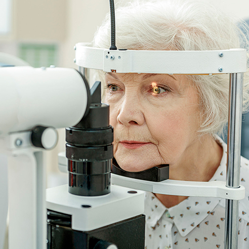
How is the eye uveitis diagnosed?
During an ophthalmological examination, the doctor reviews the patient’s medical history and performs a complex examination. This includes a series of investigations:
- Response to light;
- Blood tests;
- Imaging tests, X-ray, CT, MRI;
- Examination of the front of the eye, using a slit-lamp – to observe the optic disc, retina and its vessels, and to distinguish microscopic inflammatory cells;
- Ophthalmoscopy – to examine the back of the eye;
- Photographing the inside of the eye and retina;
- Tonometry – to measure eye pressure;
- Optical coherence tomography measuring the thickness of the retina and choroid – to determine the degree of inflammation;
- Fluorescein angiography or indocyanine green angiography – to capture an image of the blood vessels inside the eyes and see how inflamed they are;
- Examination of aqueous or vitreous fluid in the eye.
How is uveitis treated?
Depending on the severity of the disease, the treatment regimen may consist of medication or surgery. Classic treatment includes:
- systemic and cycloplegic-mydriatic medication
- or topical corticosteroids.
Infectious uveitis requires antimicrobial therapy and chronic cases require systemic corticosteroids, non-steroidal immunosuppressants, cryotherapy or surgery.
The role of treatment is:
- To reduce inflammation (with steroidal anti-inflammatories);
- To annihilate the infection (with antibiotics or antiparasitic therapy);
- To suppress the immune system (in the case of systemic autoimmune conditions, with specific drugs administered by a rheumatologist).
- Anti-inflammatory medicines;
- Medicines that control spasms;
- Medicines against bacteria and viruses;
- Medicines for the immune system;
- Drops to reduce inflammation;
- Periocular steroid anti-inflammatory injections.
- If uveitis occurs as a side effect of another disease, surgery for that disease (cataract, retinal detachment, etc.) is recommended;
- Vitrectomy – removal of the vitreous by surgery (only in severe cases)
- Implant with medicinal substance. In patients with posterior uveitis, a device may be inserted to release corticosteroids into the eye for 2-3 years.
Herbal remedies for uveitis
There are a few things that can be done at home to alleviate the symptoms of uveitis, but these cannot and should not replace expert opinion and specialist treatment.
- Applying hot or cold compresses – to relieve inflammation and eye pain;
- Wearing sunglasses – for eyes sensitive to light;
- Eating foods rich in vitamin C, E;
- Omega 3 and lutein supplementation;
- Eating foods rich in antioxidants (blueberries, salads, tomatoes, peppers, etc.).
What can you do to prevent uveitis?
- Make sure you have good eye health with proper eye and hand hygiene.
- Don’t postpone treating ophthalmological disorders and don’t hesitate to see a specialist if you have symptoms that affect your visual acuity.
The Dr. Holhoș ophthalmology network awaits you in 5 clinics across the country, with experts and state-of-the-art technology. Make an appointment and take care of your eye health!

Nystagmus is the eye condition where the eyes make repetitive and uncontrollable movements. Discover other symptoms and treatment options.

Color vision deficiency, also known as dyschromatopsia, is a general term referring to various vision disorders characterized by a deficiency in color perception.

“Flying flies” are most often harmless and represent a normal stage in the aging process. Find out what the causes are and how you can reduce the symptoms.

Ocular allergies occur as a reaction of the body to an allergen, causing inflammation and itching in the eyes. The most common ocular allergies are seasonal.

Ophthalmic migraine is most common in the 40s. It manifests itself in visual impairment and even temporary blindness.

Keratitis, also known as “corneal ulcer”, is an inflammation of the cornea. If detected early, the ophthalmological disorder is easy to treat and heals quickly.
Diplopia is an ophthalmological disease in which you see two images of the same thing. The condition can affect anyone, but is more common after the age of 60.

Xanthelasma is a member of the xanthomas family and represents fatty deposits in the skin cells around the eyes. It is visible as yellow, harmless bumps.

Colorblind people perceive colors differently from most people. Most of the time, this ophthalmological disorder makes it difficult to distinguish between certain colors.

Epiphora is an ophthalmological disorder manifested by excessive tearing of the eyes. Most of the time, it is not severe and disappears on its own. However, if you are experiencing this and the problem persists, we recommend that you make an appointment for an ophthalmological examination. Treatment can be different, depending on the cause of the epiphora.

If you notice a yellow spot on the white of your eye, you are most likely dealing with pinguecula. The ophthalmological disorder is not severe, but the symptoms can be uncomfortable. Find out how to treat pinguecula and, more importantly, how you can prevent it.

Entropion is the ophthalmological disorder in which the eyelid of the eye turns inwards. It is different from ectropion, where the eyelid turns outwards. It most often occurs in older people and usually only affects the lower eyelid.

It is possible that you may also be experiencing ocular toxoplasmosis without knowing it. This retinal disorder is extremely common in our century and is manifested by eye discomfort and blurred vision.

Ectropion is the ophthalmic disorder in which the eyelid and eyelashes pull away from the cornea, and reorient outwards.

One of the most common types of headache is headache of ocular origin. It occurs as a result of an ophthalmological disorder.

Blepharitis is an ophthalmological disorder that manifests itself by inflammation of the eyelids. At the base of the eyelids, the patient notices small crusts formed by solidified oil particles or bacteria that collect in the crease at the corner of the eye.

Uveitis is an ophthalmological disorder of the uveal tract, manifested by changes in vision and eye pain.

Among the most common ophthalmological disorders is hordeolum. This is popularly known as an “stye” and is an infection of the eyelids.

The drooping eyelid is known in medical terms as “palpebral ptosis”. It manifests itself by narrowing the visual slit of one or both eyes, creating aesthetic and functional discomfort.

Amblyopia is a vision problem, popularly known as “lazy eye”. This disorder can occur in one or both eyes, and studies show that around 3% of the population suffer from this eye disease.

The sensation of “dry eyes” or “tired eyes” is known in medical terms as “dry keratoconjunctivitis” or “xerophthalmia”, and refers to a dysfunction of the tear film.

Strabismus, also known as “crossed-eyes” or “crossed vision”, is an ophthalmological disorder in which the visual axis of the eyes is not aligned. This causes one eye to deviate when it needs to look at a fixed point.

Conjunctivitis is one of the most common ophthalmological disorders. It can occur in adults, children and babies.

Chalazion is manifested by inflammation of the upper or lower eyelid. It is one of the most common ophthalmological disorders, and occurs when the secretion of sebaceous glands in the eye is blocked.

Macular degeneration involves deterioration of the macula and therefore of the quality of central vision. This disease does not affect peripheral vision and therefore cannot lead to complete blindness.

Hypermetropia affects the ability to see nearby objects. You may be able to see distant objects clearly, but closer objects, even words in a book, are usually out of focus. Hypermetropia occurs when the eye does not focus light properly on the retina (the light-sensitive layer at the back of the eye).

Myopia is a disorder that falls into the category of refractive errors (along with astigmatism and hypermetropia). In common terms, myopia manifests itself as blurred distance vision, while near vision is not a problem.

Astigmatism, like myopia and hypermetropia, is a refractive error. In general terms, the disorder manifests itself in blurred, fuzzy vision, regardless of the distance to objects, surfaces.

Presbyopia is an age-related disorder characterized by decreased near vision. It usually appears around the age of 40.

Cataract is a common ophthalmological disorder that causes progressive loss of vision through loss of lens transparency. Studies show that about 50% of the population loses their vision due to this disorder.
Cataract is a fairly common eye disorder that causes a progressive loss of vision due to loss of lens transparency.

Diabetic retinopathy is a complication of diabetes that manifests itself at the eyes level, caused by high blood sugar levels and damage to the blood vessels of the light-sensitive tissue at the back of the eye (retina).

The retina is the light-sensitive layer deep inside the eyeball, lining the back of the eye. It is a piece of neural tissue that creates a focused two-dimensional image that is translated into a neural electrical impulse that translates the image to create visual perception.

Epiretinal membrane is a disorder of the interface area between the posterior vitreous and the central area of the retina, the macula.

The vitreous body is located in the center of the eyeball, between the lens and the retina, and appears as a “gel”. A healthy vitreous is completely transparent allowing light to reach the retina without any problems, thus obtaining a clear vision.

Glaucoma is a chronic, bilateral eye disease characterized by progressive destruction of the fibers of the optic nerve, the nerve responsible for transmitting information from the eye to the brain.

Keratoconus is a degenerative disease that manifests itself by progressive deformation and thinning of the cornea. It usually appears in adolescence, affects both men and women, and progresses into adulthood.